Female Reproductive Anatomy
The mare’s reproductive tract lies in a horizontal position within the abdominal and pelvic cavities. It includes the vulva, vagina, cervix, uterus, oviducts and ovaries (Figure 1). Changes in the anatomy or interruption in the function of any section can contribute to reproductive problems.
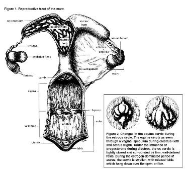
Figure 1: Reproductive Tract of the Mare
Vulva
The vulva is the exterior opening to the reproductive canal. It consists of the labia, clitoris and the vestibule. The construction of this region is important because it serves to protect the mare from the entrance of air and other contaminants into the vaginal vault.
Vagina
The vagina consists of a 6- to 8inch long muscular, mucus membrane lined tube which connects the vestibule of the vulva to the cervix. The vaginal tissues must be extremely elastic and distensible to accommodate the penis in breeding and the foal during birth.
Cervix
Basically a highly distensible muscle, the cervix is approximately 4 inches long and appears as a circle of folded tissue at the anterior surface of the vaginal vault. Its shape and characteristics change significantly in response to the body’s hormonal environment. In response to increased estrogen produced during estrus, the cervix appears pink due to increased vascularity. During this period, it produces thin, watery mucus and is so relaxed that it is often found lying limp on the vaginal floor. This flaccid cervical tone facilitates passage of semen during live cover or breeding instruments with artificial breeding. In contrast, when the cervix is under the influence of progesterone during the diestrous period and pregnancy, it produces a thick, sticky mucus, and is tightly closed and held in the center of the vaginal wall. The physical barrier produced by a healthy cervix provides a major line of defense against uterine contamination and infection. Consequently, damage to this structure can result in significant problems in maintaining fertility (Figure 2).
Uterus
This is a multi-layered, hollow, Y-shaped organ. The base of the Y is called the uterine body, while the two branches are called the horns. The uterus is suspended within the body cavity by two tough, sheet-like structures called the broad ligaments. Sagging of these ligaments with age, parity or trauma can cause a downward tilting of the uterus. This conformation can predispose the mare to the backwash of urine (urine pooling) into the reproductive tract and its accumulation at the cervix. Urine pooling can cause uterine infection and poor fertility.
The uterus is composed of three distinct layers. The outermost, serous layer is continuous with their broad ligaments. The middle layer, myometrium, consists of two sheets of muscular tissue, one oriented longitudinally and one circularly. The myometrium is responsible for the powerful contractions which expel the foal at birth. The endometrium is the innermost layer. It is a complex mucosal membrane containing a rich blood supply and many glands.
The ultimate function of the uterus is to protect, nourish and provide an environment conducive to the development of the embryo and fetus, and to expel the fetus during birth. Maintaining healthy tissues within the endometrium is crucial for optimal fertility. In fact, endometritis (uterine infection) is a major cause of infertility in mares.